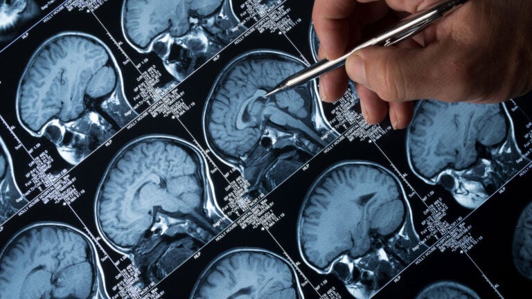
Radiofrequency ablation is a way of treating brain tumors without chemotherapy or surgery. (Photo/iStock)
USC Viterbi researchers help doctors burn away brain tumors with radio-frequency waves
USC Viterbi researchers are making it easier, faster and safer for doctors to use an emerging procedure in cancer patients
Nearly 80,000 Americans will be diagnosed with a brain tumor this year, according to the American Brain Tumor Association. Many will need major surgery and chemotherapy. Sixteen thousand will lose the battle. But USC Viterbi School of Engineering researchers are now making it easier, faster and safer for doctors to use an emerging procedure — one that uses radio-frequency waves to burn away brain tumors.
Radiofrequency ablation, or RFA, is a minimally invasive procedure that uses electrical energy to destroy cancer cells with heat. A needle-thin probe delivers radio-frequency waves directly to the tumor, cooking the tissue up to 140 degrees Fahrenheit (60 degrees Celsius) until it’s destroyed.
Monitoring and treating brain tumors
“Although ablation is becoming increasingly popular, there is still no thermal imaging technology in regular clinical use to monitor these procedures in real time and ensure that the correct thermal dose is delivered the first time,” said Research Assistant Professor John Stang of the Ming Hsieh Department of Electrical Engineering, who co-authored the study published in IEEE Transactions on Biomedical Engineering.
Together with Mahta Moghaddam, director of the Microwave Systems, Sensors, and Imaging Lab, or MiXIL, and holder of the William M. Hogue Professorship in Electrical Engineering, Stang has developed a real-time thermal imaging method and device that will help doctors deliver fast, safe and precise thermal ablation treatments for ailments ranging from tumors to epilepsy.
Surgeons and interventional radiologists rely on the guidance provided by ultrasound, CT or MRI to perform these life-saving operations. But since there is no real-time monitoring, a follow-up imaging study is needed to confirm proper treatment. This extends time in the operating room, increasing risks and costs, Moghaddam said.
“Without real-time monitoring, there is the potential for both under-treatment and over-treatment,” she said. “If there is under-treatment, doctors must perform additional rounds of thermal ablation until all of the tumor is destroyed. Each repeat ablation carries increased risk of infection or other complications and takes up more time in the operating room.”
In the case of over-treatment, there is a risk of collateral damage to the surrounding healthy tissue. This can be especially dangerous when the tumor is located close to sensitive structures, near a blood vessel or deep in the skull.
With our technology, we can guide the treatment and focus on a very specific area.
John Stang
“With our technology, however, we can guide the treatment and focus on a very specific area,” Stang said. “A microwave antenna array is placed around the region to be treated, with room left open for the surgeon to insert an ablation probe.”
Giving doctors a live temperature map
During the procedure, microwave signals are continuously transmitted and received into the treatment area. From these signals and information obtained from a prior imaging study, like an MRI, Moghaddam and Stang produce a 3-D thermal image of the region in real time, giving doctors a quantitative temperature map of the region they’re operating on.
“In in vitro experimental validation studies, our system was able to achieve one-degree Celsius accuracy at a refresh rate of one frame per second,” Stang said.
One issue the researchers have to contend with is that the resolution of their thermal image is not as high as that of MRI. But Stang sees a world in which this real-time thermal image feed can be overlaid on a high-resolution MRI, allowing doctors to precisely deliver the right dose to the right location, without the need for follow-up imaging studies.
For the next phase, their procedure will undergo animal testing later this year, specifically looking at liver cancer with support from the USC Alfred E. Mann Institute for Biomedical Engineering and in collaboration with the Keck School of Medicine of USC.
“Assuming we get good results, we may be three to five years away from clinical trials,” said Moghaddam, who last year was taking radar measurements in Alaska to map climate change in the Arctic from 40,000 feet in the air.
“This time, our environment is the human body and we make maps which are smaller,” she said. “It’s a microcosm of the larger terrestrial picture.”
Postdoctoral researchers at MiXIL Guanbo Chen and Pratik Shah contributed to this work.