
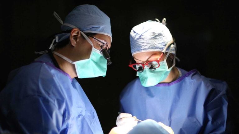
Pediatric ocular oncologists Jonathan Kim, left, and Jesse Berry collaborated on developing a liquid biopsy for children with retinoblastoma. (Photo/Thomas Lee)
Retinoblastoma study suggests way to diagnose tumor
Research at USC Roski Eye Institute and Children’s Hospital Los Angeles focuses on a liquid biopsy
Retinoblastoma, a tumor of the eye that affects children, may result in loss of one or both eyes if not diagnosed early. Although this type of tumor cannot be directly biopsied, investigators at Children’s Hospital Los Angeles and USC may have found a way to get genetic information from a tumor without removing the eye.
Results of the study by a team of investigators at the Vision Center of Children’s Hospital Los Angeles and the USC Roski Eye Institute, part of Keck Medicine of USC, were published in JAMA Ophthalmology on Oct. 12.
Genetic origin
Retinoblastoma was one of the first tumors to have its genetic origin identified; the RB1 retinoblastoma tumor suppressor gene mutation was discovered by A. Linn Murphree, a co-author on this paper, who established the Retinoblastoma Program at Children’s Hospital Los Angeles. However, ocular oncologists have been limited in their ability to use this genetic information to inform diagnosis and the application of personalized treatments— because removing tissue from the tumor in the back of the eye could spread tumor cells outside of the eye or even to the rest of the body, resulting in a far worse prognosis for the patient.
Retinoblastoma is treated using chemotherapy given either intravenously or through an artery that goes to the eye. There are limits, however, to the amount of drug that actually reaches the eye. As a result, relapse does occur due to small tumor particles that break off — or seed — from the main tumor.
The treatment for these seeds changed dramatically in 2012 when intraocular injections of chemotherapy were shown to be safe and effective. To inject chemotherapy directly into the eye, it is first necessary to remove a small amount of fluid, called aqueous humor, from the front of the eye, to decrease the pressure within the eye prior to injection of the medication.
Previously, this fluid was simply dispensed after the procedure.
“Just as I was discarding the aqueous humor, I wondered if there was a possibility it contained tumor-derived genetic material we could use to better treat our patients,” said Jesse Berry, ocular oncologist at CHLA, assistant professor of clinical ophthalmology at the USC Roski Eye Institute and first author on the study. “In fact, we found measurable amounts of tumor DNA — genetic information from the tumor that had previously been completely unavailable from an intact eye.”
James Hicks, professor of biologic sciences at the USC Michelson Center for Convergent Bioscience and professor at the Keck School of Medicine at USC, said that the chromosomal changes in the DNA found in the aqueous humor parallel chromosomal changes found in the retinoblastoma tumor. “These findings provide proof of principle that the aqueous humor can be used for a surrogate ‘liquid’ tumor biopsy,” he said.
Significant findings
The study reported on six samples from three eyes affected with retinoblastoma, among children younger than 3 years old. Two of the eyes had been removed primarily for treatment of the disease; the third eye was receiving intraocular injections as therapy but ultimately had to be removed due to disease recurrence. Aqueous humor was taken from all three eyes and allowed investigators to compare tumor DNA in the aqueous humor to DNA found in the retinoblastoma tumor.
“Until now, we could only do genetic analysis — and base therapy on the specific pathologic tumor features — when there was no longer a possibility of saving the eye,” said Thomas C. Lee, director of the Vision Center at CHLA and associate professor at the USC Roski Eye Institute. “In the future, we hope to have the capability to specifically target therapy to the type of tumor and can anticipate better outcomes for children with retinoblastoma.”
The team of investigators plans future studies to compare tumor DNA from eyes that have been saved to those that need to be removed due to tumor recurrence.
This research has the potential to completely transform how we treat children with retinoblastoma.
Jonathan Kim
“This research has the potential to completely transform how we treat children with retinoblastoma,” said Jonathan W. Kim, director of the Retinoblastoma Program at CHLA and also director of the ocular oncology service at the USC Roski Eye Institute. “This is one of the most significant findings in retinoblastoma research in the past 20 years.”
Additional contributors to the study include Jonathan W. Kim, director of the Retinoblastoma Program at CHLA, Subramanian Krishnan, Kevin Stachelek, Emily Zolfaghari and Kathleen McGovern of the Vision Center at CHLA; and Liya Xu, Anders Carlsson and Peter Kuhn of USC.
This work was funded by Bright Eyes, the Nautica Foundation and the Knights Templar Eye Foundation. Additional support was provided by Retinoblastoma International Inc; the Larry and Celia Moh Foundation; the Institute for Families Inc, Children’s Hospital Los Angeles; Research to Prevent Blindness; the Vicky Joseph Research Fund; the Carol Vassiliadis Research Fund; and the USC Dornsife College of Letters, Arts and Sciences.